News & Events

By Dan Lewis Foundation
•
April 2, 2025
For the third consecutive year, the Dan Lewis Foundation for Brain Regeneration is proud to announce the DLF Prize competition. The 2025 DLF Prize, a $20,000 award, will recognize an outstanding early career scientist (2 to 5 years post-doc) conducting innovative research in neuroscience, pharmacology, or biotechnology. This prestigious prize honors researchers whose work aligns with the DLF mission to drive breakthroughs in neural regeneration and repair. The current research priorities of the DLF are: Pharmacological Reactivation of Neural Repair: Research into pharmacological methods of reactivating or augmenting synaptogenesis, neurogenesis or axonal repair. Cell-Based Cortical Repair: Investigating the potential of derived cortical neurons to restore function in damaged cortical regions. Transcriptomics of Neural Recovery: Characterizing transcriptomic profiles of cortical neurons in the recovery phase following brain injury to identify pathways that drive repair. Molecular Inhibitor Targeting: Advancing anti-sense oligonucleotides (ASO’s) or small-molecule therapeutics designed to downregulate inhibitors of neural regeneration in the cortex or spinal cord. Application for the 2025 DLF Prize can be made by going to our website— danlewisfoundation.org —and clicking on the Tab “ 2025 DLF Prize ”. This will bring you into the application portal. The application portal opened in March, 2025 and will remain open through May 31st. Once in the portal, you will find complete information about the DLF prize, eligibility requirements, and an application form which can be filled in and submitted online. The winner of the 2023 DLF Prize, Dr. Roy Maimon, continues his research indicating that downregulation of PTBP1, an RNA-binding protein, can convert glial cells into neurons in the adult brain (Maimon et al. 2024) .* Dr. Maimon, currently a post-doc at the University of California, San Diego is currently interviewing for a faculty position at several prominent neuroscience departments. The winner of the 2024 DLF Prize, Dr. William Zeiger is a physician-scientist in the Department of Neurology, Movement Disorders Division, at UCLA. Dr. Zeiger has expertise in interrogating neural circuits using a classic “lesional neurology” approach. He states, “Our lab remains focused on understanding how neural circuits become dysfunctional after lesions to the cortex and on investigating novel circuit-based approaches to reactivate and restore damaged cortex”. * Maimon, Roy, Carlos Chillon-Marinas, Sonia Vazquez-Sanchez, Colin Kern, Kresna Jenie, Kseniya Malukhina, Stephen Moore, et al. 2024. “Re-Activation of Neurogenic Niches in Aging Brain.” BioRxiv. https://doi.org/10.1101/2024.01.27.575940.
By Dan Lewis Foundation
•
April 2, 2025
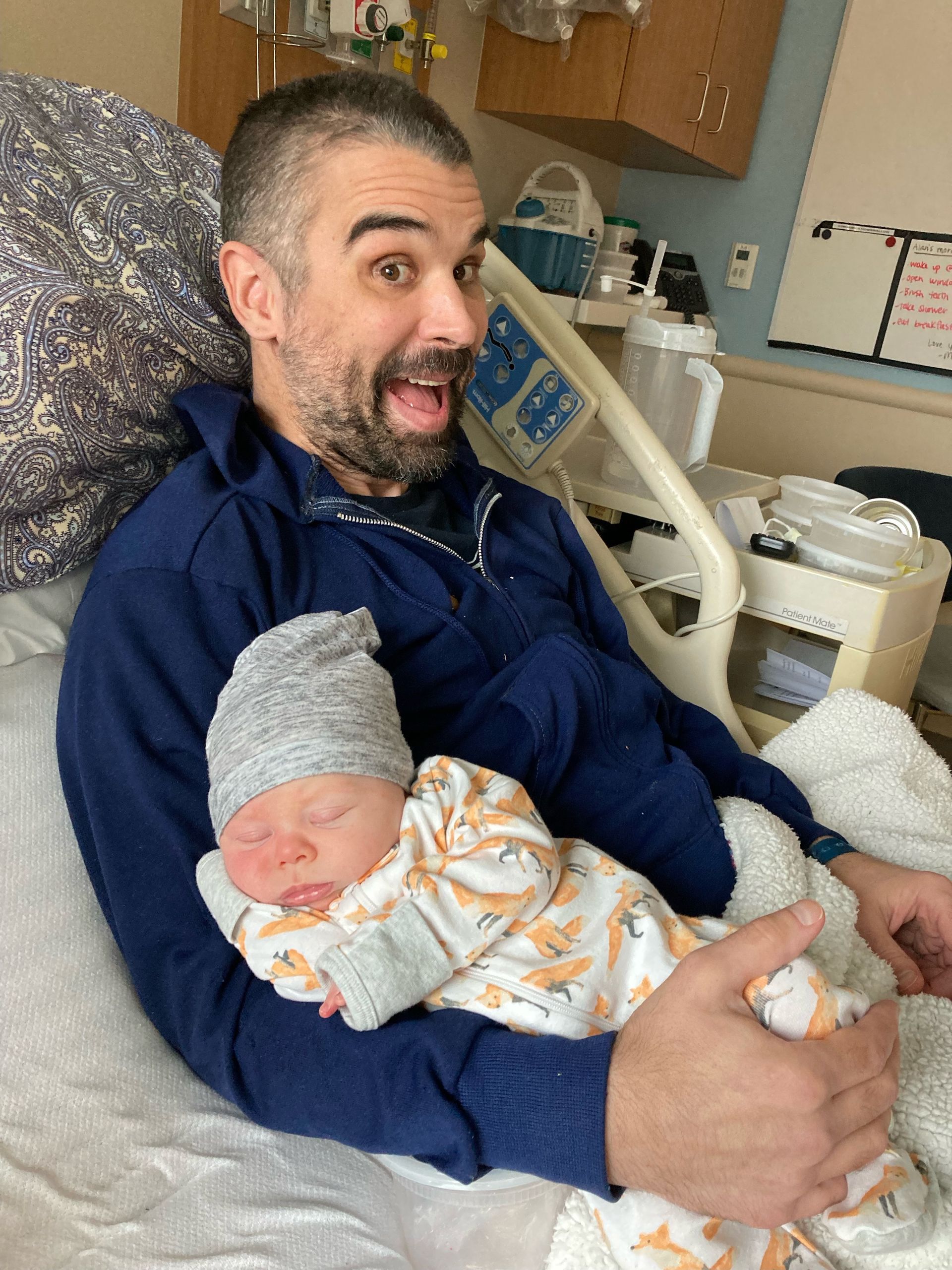
Alan was injured in 2021, at age 42. An art teacher in Lakewood, Colorado, Alan was riding his bicycle after school and was crossing at an intersection when a truck turned into the crosswalk area and hit him. Alan reports no memory of the event but has been told this is what happened. Alan says “My frontal lobe took the brunt of the impact, particularly the left frontal lobe”. Alan had a 2 ½ week stay at a nearby hospital where he, “re-learned to talk, to walk, and drink”-- although again he reports no memory of his stay there. Alan was then transferred to Craig Rehabilitation Hospital, in Englewood, Colorado. Alan says, “The only reason I knew I was at Craig is that I rolled over in bed and saw “Welcome to Craig” on the dry erase board.” During this stage of recovering, Alan repeatedly denied that he had been in an accident. Twice he tried to leave Craig on his own accord despite his wife’s and his therapists’ assurances that it was important for him to stay to recuperate from his injuries. Alan’s wife was 8 months pregnant at the time of his accident and gave birth to their son while Alan was an inpatient at Craig. Alan’s wife brought his newborn son to visit him days after the birth and Alan held him while sitting in his wheelchair, but Alan wistfully reports this is another thing he can’t remember. Alan reports that he still has significant difficulties with memory. Alan has also experienced several other neuropsychological difficulties. He states that for months after his injury, he could not experience emotion. “I could not laugh, I couldn’t cry.” Even after three years, his emotional experience is constricted. However, an emotion that is sometimes elevated is irritation and anger. Sometimes, dealing with people can be difficult because he may have temper flare-ups with little reason. This is something that Alan regrets and he is working hard with his neuropsychologist to improve the regulation of his emotions. Alan also has difficulty with organization, motivation, and distractibility. Earlier in his recovery, he had trouble sequencing and had difficulty carrying out personal and household routines. Alan has benefited greatly from therapy and his own hard work to make improvements in these areas. A chief reason that Alan works so hard in his recovery is so that he can be a good father to his son who is now almost 3 years old. He recognizes that it is important not to get frustrated when it seems that he can’t provide what his son wants or needs at a given moment. “I’m trying to raise my son the best I can…he’s at such a pivotal time in his life.” Alan’s financial situation was helped for a time by Social Security Disability Insurance payments but these payments ended. He is trying to get SSDI reinstated but the process of doing so is confusing and is taking a lot of time. Alan returned to work about 11 months ago at a liquor store (after about 2 years of not being able to work), the same store where he previously worked part time while teaching. He works in the wine department. “I sell wine and make recommendations.” When asked for advice to other brain injury survivors, Alan’s words were: “No matter how confused or upset you are or how frustrated you get, keep pressing on and moving forward because there is light at the end of the tunnel even though it may seem long. Keep moving forward and don’t give up no matter what anyone says to you”. Alan added that supports for individuals with brain injury are very important. He has found support groups, retreats, and seminars/events where brain injury survivors can share their experience to be very helpful. The volunteer work he does at Craig Hospital has been valuable for him. Alan is an inspiring individual. Despite having scarce memory of his accident and some confusion about the functional losses he has experienced, Alan has worked hard to make his recovery as complete as possible. He continues to work hard to progress and to express gratitude for those who have assisted him along the way.
By Hal C. Lewis, Ph.D. & David Margulies, M.D.
•
April 2, 2025
The overarching goal of the Dan Lewis Foundation for Brain Regeneration Research (the DLF) has been, for the past five years, to support biomedical and biotechnological discoveries that will bring the promise of regenerating and repairing the seriously damaged brain into reality. We pursue this goal by sponsoring neuroscientific conferences, publishing a quarterly newsletter, funding early career scientists with the DLF Prize program, sharing promising research nationwide, and posting brain injury information on social media. We strongly believe that biomedical and biotechnological solutions and options for regenerating the seriously injured brain are not only possible but inevitable. The key question is not an “if” question but rather a “how soon” question. Unfortunately, the current U.S. administration has adopted a somewhat negative stance towards the sciences in general, including the health sciences and biomedical research. With disregard for the way scientific knowledge accumulates, the budget slashers at DOGE, under the direction of the President and his Cabinet, have threatened serious cuts to NIH and NIMH research grants as well as to funds supporting students and faculty in neuroscience and biomedical training programs. Serious cuts to funding have already been made and further reductions can be expected. This short-sighted policy approach will significantly delay important discoveries across various scientific fields. Specifically, for DLF’s constituency, discoveries and related clinical trials that were potentially only a few years away may now be postponed by 5 to 10 years. The DLF continues to stand in support of neuroscientific and biomedical programs, their students, trainees, faculty, and researchers that have contributed so much to the field of regenerating the damaged brain. If you visit our website—danlewisfoundation.org—you will see “Donate” buttons scattered throughout the content. Please consider a donation, no matter how small or large—to help us continue our work to catalyze progress in brain regeneration research and to urge policy makers to correct course and return to solid support of biomedical and biotechnological advances that will be of so much value to the American public and to persons with serious brain injuries worldwide.
By David Margulies, M.D.
•
April 2, 2025
Introduction: The Paradigm Shift in Brain Regeneration For decades, scientists believed that the adult brain was incapable of meaningful regeneration. Unlike skin or muscle, the central nervous system (CNS) appeared to lack the ability to repair itself after injury, and lost neurons were considered irreplaceable. However, cutting-edge research now demonstrates that this is no longer the case. The brain possesses dormant repair mechanisms—pathways that were active during development but have been shut down in adulthood. By reactivating these pathways, it may be possible to induce neurogenesis (the birth of new neurons), axonal repair, and synaptogenesis (the formation of new neuronal connections) after devastating injuries like stroke, traumatic brain injury, and spinal cord damage. A growing body of evidence from both the spinal cord and the central nervous system (the CNS) shows that regeneration can be stimulated by downregulating the repressing factors that prevent neuron growth and repair. For clarity, “downregulation” means reducing the activity or number of something. In the case of a repressing factor, downregulation means lowering its levels or making it less active. A repressor is a protein that blocks a process—often by preventing a gene from being turned on. When the repressor is downregulated, it no longer effectively blocks that process, allowing the underlying function to be released or activated. These advances open the door to new treatments that could restore function to patients with neurological injuries, potentially reversing what were once thought to be permanent disabilities. The Science Behind Reactivating Brain Repair Neurodevelopmental genes and pathways that promote neuronal growth and plasticity are switched off after early development. However, researchers have identified key molecular regulators that act as “brakes” on brain regeneration. The “brakes” act as repressors of brain regeneration. By inhibiting these repressors, the brain’s intrinsic ability to regrow neurons and axons can be reactivated. Recent breakthroughs include: Neurogenesis in the Adult Brain : Research has shown that downregulation of PTBP1, an RNA-binding protein, can convert glial cells into neurons in the adult brain. Studies using antisense oligonucleotide (ASO) therapy to transiently suppress PTBP1 have successfully induced the formation of functional neurons in mouse models of neurodegeneration. For clarity, an antisense oligonucleotide (ASO) is a short strand of DNA or RNA designed to bind to a specific mRNA and regulate gene expression. By binding to its target, an ASO can block protein production, modify splicing, or mark the mRNA for destruction, effectively acting as a precise “off switch” for genes. In this context ASOs can be used to release glial cells to develop into functional neurons. Work is now underway to create ASOs that can be safely administered to humans, thereby stimulating the creation of new neurons. Axonal Regeneration in the CNS: Another major discovery involves unlocking nerve fiber regrowth by downregulating specific nerve growth inhibition, which prevents nerve fiber regrowth. Studies have demonstrated that suppressing these inhibitors leads to long-distance axonal regeneration, restoring function in spinal cord injury models. Synaptic Repair in Neurodegenerative Disease: Scientists have shown that modulating key synaptic receptors—such as mGluR5—can restore lost synaptic connectivity, improving brain network function in neurodegenerative diseases like Alzheimer’s. The neuronal connections and circuits in the brain are maintained in a stable state by the balanced action of different synaptic receptors. By modifying this balance, the rate of brain synapse formation can be increased or decreased. Together, these findings confirm that neural repair is possible when the appropriate repressive factors are removed, unlocking the brain’s natural regenerative capacity. Quiver’s Scalable Human Neuronal Platform: Accelerating Discovery While these discoveries are promising, translating them into effective therapies requires precise, scalable, and high-throughput screening technologies that can screen out or discover biomolecules that address a specific biomedical target by the thousands with great rapidity. One such technology has been developed by Quiver Biosciences using an all-optical electrophysiology platform. (Note: the author of this article is a co-founder of Quiver Biosciences) Quiver has developed a human induced pluripotent stem cell (hIPSC)-derived neuronal platform that allows researchers to measure functional activity in human neurons at an unprecedented scale. This platform is uniquely capable of: Directly measuring neuronal excitability, synaptic transmission, and network connectivity using optogenetics and advanced imaging techniques. Screening for new factors that inhibit brain repair, helping scientists identify additional repressors that need to be targeted. Evaluating small molecules and ASO-based therapies that may upregulate brain regeneration, assessing their efficacy and toxicity before advancing to clinical trials. The potential of this platform is vast. Scientists can now systematically search for drugs that mimic the effects of PTBP1 suppression, Nogo-A blockade, or mGluR5modulation—treatments that could one day be used to regrow neurons, reconnect severed axons, and restore lost synapses in patients with brain injuries. Key advantages of Quiver’s platform include: Scalability: Unlike traditional animal models, this system enables high- throughput drug screening on human neurons, increasing the efficiency of therapeutic discovery. Relevance: Because the neurons are derived from human stem cells, they offer a more accurate model of human brain function and disease than animal-based approaches. Safety Screening: The platform allows for early identification of toxic effects, ensuring that only the safest and most effective compounds move forward in development. By integrating AI and machine learning, Quiver’s platform also enables pattern recognition of successful drug candidates, identifying compounds with optimal efficacy and minimal side effects. The Role of Philanthropy: Funding the Future of Brain Repair Scientific breakthroughs alone are not enough to bring regenerative therapies to patients. Translational research—the process of developing basic discoveries into real-world treatments—is slow, expensive, and underfunded. This is where philanthropic support can make an immediate impact. Many of the pioneering studies on brain regeneration were initially considered too risky or unconventional for traditional funding sources. Yet, thanks to early philanthropic investments, these ideas have now been validated and are shaping the next generation of treatments. Today, funding is urgently needed to: Expand screening for regeneration-promoting drugs Optimize ASO-based approaches for inducing neurogenesis and axonal repair. Accelerate clinical trials for promising regenerative therapies. A New Era for Brain Regeneration For the first time, we have the tools to reactivate the brain’s own repair mechanisms, offering hope for individuals with severe neurological injuries. The path forward is clear: by investing in innovative research, scaling up discovery efforts, and supporting translational studies, we can bring brain-regenerating therapies to patients faster. The DLF raises funds and uses them to inspire, catalyze, and accelerate work towards brain regeneration. We increase public awareness of possibilities in brain regeneration through our social media, news “blasts”, and quarterly newsletter. And, we promote neuroscientific advances through consultation, networking, conferences, and seed grants. This is especially important when federal funding is limited. By supporting this work, we can help transform groundbreaking scientific insights into real treatments that restore lost function and improve lives.
By Dan Lewis Foundation
•
November 13, 2024
Stroke is a common neurological condition that damages brain cells (neurons) in the affected area, leading to a loss of the functions controlled by that region. A hopeful aspect of stroke recovery is that, over time and with rehabilitation, many individuals regain some abilities. This recovery has been linked to a process called “remapping,” where neurons in unaffected areas of the brain adapt to take over the functions of the damaged areas. Although many studies have explored this remapping phenomenon, most evidence has been indirect, based on changes in brain activation patterns or neuron connections after stroke in animal models. Direct proof that neurons change functionality after stroke has been lacking, partly because measuring neuron activity in the brain over time, especially at the necessary scale and duration, is challenging.
By Dan Lewis Foundation
•
November 6, 2024
After a life-altering accident in October 2022, Devon Guffey’s story is about resilience and determination. His journey has been profiled in the summer 2023 issue of the Making Headway Newsletter: https://www.danlewisfoundation.org/devons-story . Hit by a drunk driver, Devon sustained severe brain and physical injuries, including axonal shearing, a traumatic frontal lobe injury, and facial fractures. Even after contracting meningitis while in a coma, Devon fought hard to survive – and today, his recovery continues to inspire us all. In late 2023, Devon worked as an assistant basketball coach at Blue River Valley, where he had once been a student. His love for sports and dedication to regaining his physical strength returned him to the gym, where his hard work paid off. Devon’s persistence earned him another job at the YMCA, guiding gym members and supporting facility upkeep. Through all the challenges—deafness in one ear, blindness in one eye, and a permanent loss of taste and smell—Devon perseveres. He recently regained his driving license, a significant milestone that symbolizes his increasing independence and cognitive and physical recovery. While each day may not show significant changes, Devon now sees his progress over time. Today, Devon speaks to groups about his journey, the dangers of drunk driving, and finding strength in adversity. His message is clear: recovery is a process, and sometimes, "can't" simply means "can't do it yet ." Every TBI is unique, and Devon’s story powerfully reminds us of the strength that comes from resilience and community. We are grateful to Devon for continuing to share his story and for his role in uplifting others facing difficult paths. His journey is a testament to the fact that we are stronger together. #BrainInjuryAwareness #DevonsJourney #Resilience #EndDrunkDriving #MakingHeadway

By Dan Lewis Foundation | Summer 2024
•
July 10, 2024
Scientists worldwide are working to find ways to stimulate healing and functional recovery after severe brain injuries. This work is driven by the desperate needs of persons who have suffered brain damage. It is inspired by the knowledge that the information required to create new brain cells, cause these cells to interconnect, and stimulate new learning is contained in our genome. Now that we can readily generate stem cells from adult tissue, we have access to the genomic program that can control all of the intricate details of brain tissue formation. A number of different research themes are being pursued productively. These include: (1) enabling injured neurons to self-repair (“axonal repair”) 1,2 ; (2) replacing damaged tissue by increasing the growth of new neurons (“neurogenesis”) 3-5 ; (3) transplanting new brain cells that are derived from a person’s own stem cells (“autologous cellular repletion”) 6-8 ; (4) stimulating the re-wiring of new or surviving tissue by encouraging the formation of new connections (“synaptogenesis”) 9,10 ; and (5) augmenting the function of a damaged brain by the use of bio-computational prostheses (“brain-computer interfaces”) 11,12 ; We’ve explored these themes in previous newsletters. The goal of stimulating meaningful brain regeneration is now sufficiently plausible that a large-scale, well-funded campaign needs to be funded to bring meaningful new therapies to patients within the foreseeable future. Here, we suggest a high-level outline of the research themes for such a campaign. A ‘moon shot’ program towards brain regeneration would leverage cutting-edge technologies in stem cell research, gene therapy, synaptic plasticity, neuronal repair, and brain-computer interfaces (BCIs) to develop innovative treatments for brain injuries and neurodegenerative diseases. These treatments would target the restoration of lost brain functions and improvement in the quality of life for individuals affected by severe brain injuries. This research agenda aims to catalyze serious discussion about creating a federal program with funding, organizational resources, and expert governance to enable brain regeneration in our lifetimes. Major Themes For a Brain Regeneration “Moon Shot” Program 1: Promote the formation of new neurons 1.1 Stimulate the brain to create new neurons 1.2 Create new neurons from patient-derived induced pluripotent stem cells to be transplanted back into the patient. Create new glial cells to support neurogenesis. 2: Stimulate new synaptic formation 2.1 Develop drugs that enhance synaptic plasticity and promote the formation of new synaptic connections 3: Stimulate self-repair of damaged neurons 3.1 Develop drugs that de-repress neurons and, thereby, enable axonal regrowth 4: Develop brain-computer interfaces (BCIs) for brain-injured patients 4.1: Develop and test BCIs that enable the brain to control behaviors or external devices and, thereby, augment or replace impaired functions. 4.2: Develop and test BCIs that can accelerate the training of remapped brain tissue in persons with brain injuries to optimize functional recovery. 4.3: Combine BCIs with other strategies (e.g., cell repletion, synaptogenesis, and enhanced plasticity) to accelerate adaptation and functional improvement. The proposed research themes can underpin targeted research to stimulate meaningful brain regeneration, offering new hope for patients with brain injuries and neurodegenerative diseases. While the scientific challenges are profound, there has been sufficient progress to justify substantial investment in brain regeneration research. Any such large-scale program will require coordinated collaborations among academic and commercial partners, skillful governance and management, and a shared sense of profound commitment to the goal. The recent pace of advances in cell biology, stem cell technology, bio-computational interfaces, and genomically targeting medicines suggests that large-scale investment will yield meaningful clinical advances toward brain regeneration after injury. With robust funding and skilled leadership, this comprehensive research agenda has a realistic potential to transform scientific breakthroughs into tangible medical therapies, offering hope to millions affected by brain damage.

By Dan Lewis Foundation | Summer 2024
•
July 10, 2024
Stephen Mark Strittmatter, MD, PhD , earned his undergraduate degree from Harvard, and completed doctoral training at Johns Hopkins. His medical internship and neurology residency were at Massachusetts General Hospital. He joined the Yale faculty in 1993, and is now Chair and Professor of Neuroscience and Vincent Coates Professor of Neurology. He is Director of the Kavli Institute for Neuroscience at Yale, the Yale Cellular Neuroscience, Neurodegeneration and Repair Program, the Yale Alzheimer Disease Research Center and the Yale Memory Disorders Clinic. His work on developmental axonal guidance led to his discovery of a Nogo Receptor pathway critical for axonal re-growth after injury. He showed that glia-derived inhibitors bind this receptor, activate RhoA and prevent neural repair. He developed a soluble Nogo Receptor decoy protein that blocks the endogenous ligand-receptor interaction, promoting recovery from spinal cord injury and stroke. This therapeutic protein is now in clinical trials for patients with chronic spinal cord injury. Strittmatter has also translated discoveries in neurodegeneration to clinical approaches. Using innovative screens, he identified the roles of Prion Protein and metabotropic glutamate receptor 5 (mGluR5) as Aß oligomer receptors. He linked activation of these receptors to a pair of synaptic tyrosine kinases, the Tau-interacting Fyn and the AD risk gene PTK2B. Critically, this pathway contributes to synapse loss and memory deficits in preclinical models. He subsequently identified a Fyn inhibitor which was tested in an AD clinical trial. Strittmatter has developed additional methods for targeting this pathway with robust preclinical efficacy. One approach uses novel mGluR5 silent allosteric modulators, which are now in clinical trials. An author of over 280 original reports, Dr. Strittmatter has been recognized by the King Faisal International Prize in Medicine, Ameritec Award for Spinal Injury Research, McKnight Brain and Memory Disorders Award, Alzheimer Association Zenith Fellow Award, Senator Jacob Javits Award in the Neurosciences, and an NINDS Outstanding Investigator Award.

By Dan Lewis Foundation | Summer 2024
•
July 10, 2024
Towards Brain Regeneration and Functional Recovery A significant brain injury can result in the loss of brain tissue, disruption of nervous system connections, destruction of brain regions controlling various functions, and chaotic biochemical and electrical activity. To minimize the initial injury’s impact, it is important to control bleeding and swelling, limit ongoing damage and scarring, and limit harmful cascading metabolic processes. Human brain tissue does not regenerate, but some functional recovery can occur as surviving brain regions retrain to take over lost functions. Survivors of severe brain injury often experience only modest improvement over time. Recent research has indicated that developing biomolecular medicines that can stimulate brain regeneration and functional recovery is plausible, even years after the injury. The DLF is dedicated to supporting the creation of biomedical therapies for brain regeneration for persons with severe brain damage. Their strategies include repletion of neurons, enhancement of synaptic connections, reconnection of severed axons, targeted training of brain regions, and innovative use of brain-computer interfaces. The DLF newsletter has and will continue to showcase cutting-edge research to inspire support for this important work. The DLF relies on charitable contributions and grants to fund research. Every donation, whether large or small, helps to make our vision of meaningful brain regeneration and improved functional outcomes closer to reality. To learn more: https://www.danlewisfoundation.org/towards-brain-regeneration-and-functional-recovery The Synapse and Brain Regeneration The brain largely consists of interconnected neurons carrying electrical currents. These currents are transmitted by neurotransmitters at a microscopically small gap (the synapse) between neurons. Neurotransmitters carry the signal across the synapse, thus allowing the signal to pass from one neuron to the next. The brain relies on trillions of these synaptic connections, which form established pathways but also allow the brain to alter itself in response to stimulation to adapt to new experiences. Recent breakthroughs in research have significantly advanced our understanding of synaptic formation, neurotransmitter roles, and the effects of drugs on cross-synaptic communication. After severe brain injury, the formation of new synaptic connections is crucial for functional recovery. Studies have shown that stimulating the connection between neurons is a highly promising strategy for brain regeneration. Another innovative approach involves replacing lost neurons by stimulating new growth or transplanting cells. Providing a rich environment that supports synaptic connections through retraining helps promote neuroplasticity. To learn more: https://www.danlewisfoundation.org/the-synapse-and-brain-regeneration Can Damaged Brain Tissue Be Replaced? Brain regeneration research, a novel and cutting-edge area, is dedicated to the regrowth or regeneration of brain tissue. Recent advances involve creating induced pluripotent stem cells (iPSCs) from skin cells, which can form any mature tissue, including nerve tissue. Researchers hope to restore lost brain functions by reintroducing these stem cells into damaged brains. For instance, iPSCs can generate dopamine-producing neurons to replace those lost in Parkinson’s disease. Studies in monkeys show that transplanted iPSC-derived neurons can survive, integrate, and improve motor function, with promising results in human clinical trials. Despite progress, many scientific, technical, and ethical challenges remain. However, cultivating induced pluripotent stem cells from an individual’s own existing cells offers significant hope for meaningful regeneration of the injured brain and better recovery. To learn more: https://www.danlewisfoundation.org/can-damaged-brain-tissue-be-replaced Targeting the Genome to Promote Brain Regeneration The entire set of DNA instructions in a cell (the genome) directs the brain’s development, growth, and maturation. DNA, containing the genetic code unique to each individual, is encoded into a similar molecule called RNA. The RNA then carries genetic information that is translated into various proteins necessary for neuronal proliferation and health Researchers are exploring several methods to stimulate brain regeneration at the genetic level: Gene Therapy: This method introduces new genetic material into adult cells using an engineered “de-activated” virus to carry DNA to target cells to promote neuronal repletion. This method has shown promise in terms of the production of proteins necessary to protect existing neurons or to regenerate neurons in neurodegenerative diseases. Gene Editing: Techniques like CRISPR-Cas9 enable precise genomic corrections. Successful trials for genetic blindness offer hope for similar applications in brain regeneration. Antisense Oligonucleotides (ASOs): These are small synthetic chains of amino acids that bind to RNA molecules to modulate gene activity. ASOs can inhibit the production of proteins harmful to neurological development or promote the production of beneficial proteins, showing great promise in diseases like spinal muscular atrophy, Huntington’s disease, and ALS. To learn more: https://www.danlewisfoundation.org/targeting-the-genome-to-promote-brain-regeneration Unlocking the Regenerative Powers of Antisense Oligonucleotides for Brain Injury Recovery The brain’s inherent limitations in regeneration pose significant challenges in the recovery from brain injuries and neurological disorders like Alzheimer’s and Parkinson’s. Recent strides in molecular biology and genetics, particularly with antisense oligonucleotides (ASOs), hold immense promise for novel and effective treatments. ASOs interact with RNA to block gene expression, potentially enhancing regeneration by: Promoting neurogenesis by targeting genes that regulate neuron formation. Reducing inflammation by silencing inflammatory process genes. Enhancing axon (portion of the nerve cell that sends signals at the synapse) regrowth to re-establish functional connections. Challenges for ASO therapies include ensuring specificity to avoid off-target effects and ensuring effective delivery across the blood-brain barrier. Nevertheless, ASOs represent a very exciting path towards brain regeneration. To learn more: https://www.danlewisfoundation.org/unlocking-the-regenerative-powers-of-antisense-oligonucleotides-for-brain-injury-recovery Brain Regeneration via Brain Tissue Transplantation: A Glimpse into the Future of Medicine Replacing a severely damaged liver with a healthy portion is possible; replacing brain tissue is far more challenging. The first major hurdle in brain tissue transplantation is sourcing replacement brain tissue. With recent breakthroughs, scientists are now able to transform readily available blood or skin cells into pluripotent stem cells (iPSCs) and reprogram them into neurons. When cultured, these neurons can mimic intact brain neurons and are not rejected as foreign tissue when transplanted back into the individual. A decade ago, scientists successfully grew derived neurons into organoids -- small ‘mini-brains’ with many features of a living brain. Despite limited survival in cell cultures, these organoids demonstrated that derived neurons possess all the necessary information to create a partially functional brain in the laboratory setting. With their ability to develop new neural connections, organoids hold immense potential in compensating for damaged brain tissue and promoting recovery. However, their application in humans necessitates the creation of suitable transplantation sites, developing surgical techniques, and optimizing tissue integration without disrupting brain activity. To learn more: https://www.danlewisfoundation.org/brain-regeneration-via-brain-tissue-transplantation Brain-Computer Interfaces to Augment Brain Regeneration A Brain-Computer Interface (BCI) enables direct communication between the brain and external devices, allowing control through thought. BCIs facilitate actions like typing, playing music, controlling prosthetics, or steering wheelchairs by thinking. They can also reconnect brain regions to the body or external world after neuronal connections are lost, providing sensory input or motor output. How Do BCIs Work? BCIs are not just about decoding and encoding the brain’s electrical signals. They are a complex interplay of technology and biology. BCIs use sensors placed on the scalp (non-invasive) or within the brain (invasive) to detect these signals. Sophisticated algorithms are the key to making BCIs work. These algorithms interpret the signals, enabling control of prosthetic limbs, cursors, or other devices and transmitting sensory information directly to the brain. BCIs hold immense potential to transform the lives of individuals with severe brain injuries. They can empower paralyzed individuals to regain control over their limbs, expedite brain reprogramming, and enable the use of external devices. Biologic Augmentation of BCI Benefits New medicines that stimulate neuron formation, repair damaged axons, and enhance synaptic connections have the potential to amplify the benefits of brain-computer interfaces. These treatments might aid BCI recipients by preconditioning the brain or replacing lost tissue. However, challenges remain, including ethical considerations, technological limitations, and the need for personalized rehabilitation. Despite these hurdles, BCI technology shows promising potential for restoring abilities to those with severe brain injuries. To learn more: https://www.danlewisfoundation.org/brain-computer-interfaces-to-augment-brain-regeneration
Your support can make a difference
Please consider donating. Every dollar donated goes toward keeping our research alive.
The Dan Lewis Foundation for Brain Regeneration Research is an incorporated 501(c)(3) non-profit organization. Donations are tax deductible.
Thank you to our sponsors: Google for Non-Profits
Donations of stock may have significant tax benefits for the donor and are a great way to support the foundation. Please contact us for more information regarding the tax benefits and the process of transferring stock to the foundation.
Subscribe for Updates
Subscribe to our newsletter
Thank you for contacting us.
We will get back to you as soon as possible.
We will get back to you as soon as possible.
Oops, there was an error sending your message.
Please try again later.
Please try again later.
© 2025
All Rights Reserved | The Dan Lewis Foundation For Brain Regeneration Research | In Partnership With CCC
